Figure 1. [The normal human retina fundus]. - Webvision - NCBI
Por um escritor misterioso
Descrição
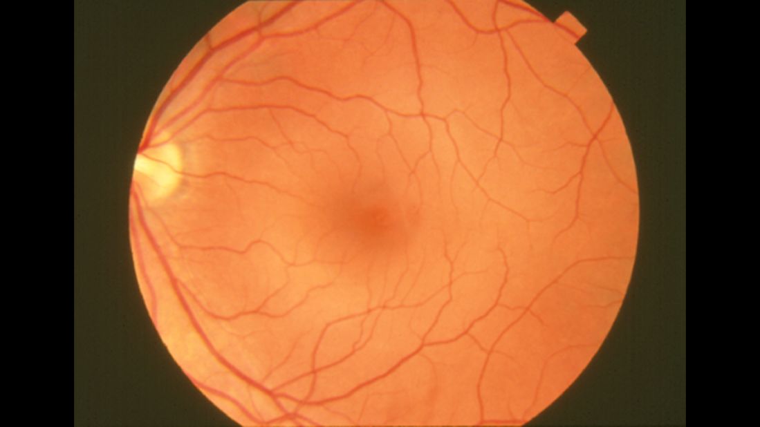
The normal human retina fundus photo shows the optic nerve (right), blood vessels and the position of the fovea (center).
![Figure 1. [The normal human retina fundus]. - Webvision - NCBI](https://media.springernature.com/lw685/springer-static/image/chp%3A10.1007%2F978-3-030-25886-3_22/MediaObjects/436773_1_En_22_Fig7_HTML.png)
Image Analysis for Ophthalmology: Segmentation and Quantification of Retinal Vascular Systems
![Figure 1. [The normal human retina fundus]. - Webvision - NCBI](http://webvision.med.utah.edu/wp-content/uploads/2015/10/ArdenFig4new.jpg)
Diabetic Retinopathy and A Novel Treatment Based On The Biophysics Of Rod Photoreceptors And Dark Adaptation by Geoffrey. B. Arden and David J. Ramsey – Webvision
![Figure 1. [The normal human retina fundus]. - Webvision - NCBI](https://www.cell.com/cms/asset/caee552e-ec98-489a-bfee-13674a9775ca/fx1.jpg)
Cell therapy with hiPSC-derived RPE cells and RPCs prevents visual function loss in a rat model of retinal degeneration: Molecular Therapy - Methods & Clinical Development
![Figure 1. [The normal human retina fundus]. - Webvision - NCBI](https://www.ncbi.nlm.nih.gov/books/NBK543075/bin/diseases_glaucoma-Image005.jpg)
Figure 4. [The course of ganglion cell]. - Webvision - NCBI Bookshelf
![Figure 1. [The normal human retina fundus]. - Webvision - NCBI](https://media.springernature.com/full/springer-static/image/art%3A10.1038%2Fs41598-022-22964-w/MediaObjects/41598_2022_22964_Fig1_HTML.jpg)
Effective field of view of wide-field fundus photography in the Stanford University Network for Diagnosis of Retinopathy of Prematurity (SUNDROP)
![Figure 1. [The normal human retina fundus]. - Webvision - NCBI](https://www.mdpi.com/applsci/applsci-08-00155/article_deploy/html/images/applsci-08-00155-g001.png)
Applied Sciences, Free Full-Text
![Figure 1. [The normal human retina fundus]. - Webvision - NCBI](https://webvision.med.utah.edu/wp-content/uploads/2019/07/KrizajFigure4sure.jpg)
What is glaucoma? by David Krizaj – Webvision
![Figure 1. [The normal human retina fundus]. - Webvision - NCBI](https://www.ncbi.nlm.nih.gov/books/NBK470669/bin/myopia-Image005.jpg)
Figure 3. [A fundus photograph of the]. - Webvision - NCBI Bookshelf
![Figure 1. [The normal human retina fundus]. - Webvision - NCBI](https://www.ncbi.nlm.nih.gov/books/NBK554706/bin/Archetecture_Fovea-Image012.gif)
The Architecture of the Human Fovea - Webvision - NCBI Bookshelf
![Figure 1. [The normal human retina fundus]. - Webvision - NCBI](https://journals.physiology.org/cms/10.1152/physrev.00035.2019/asset/images/medium/z9j004202952r001.png)
Emerging Approaches for Restoration of Hearing and Vision
![Figure 1. [The normal human retina fundus]. - Webvision - NCBI](http://eyerounds.org/atlas/LARGE/Normal-fundus-LRG.jpg)
Atlas Entry - Situs Inversus of the Retinal Vessels
![Figure 1. [The normal human retina fundus]. - Webvision - NCBI](https://journals.sagepub.com/cms/10.1177/15353702211022674/asset/images/large/10.1177_15353702211022674-fig1.jpeg)
Interpretation of anatomic correlates of outer retinal bands in optical coherence tomography - Xincheng Yao, Taeyoon Son, Tae-Hoon Kim, David Le, 2021
![Figure 1. [The normal human retina fundus]. - Webvision - NCBI](https://ars.els-cdn.com/content/image/1-s2.0-S266646902300026X-gr1.jpg)
Cell death mechanisms in retinal phototoxicity - ScienceDirect
de
por adulto (o preço varia de acordo com o tamanho do grupo)