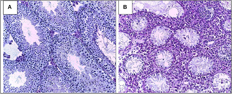
Morphology of Leydig cells in the testes after in vivo MCP-1 treatment.
Por um escritor misterioso
Descrição
Frontiers Pathomechanisms of Autoimmune Based Testicular Inflammation
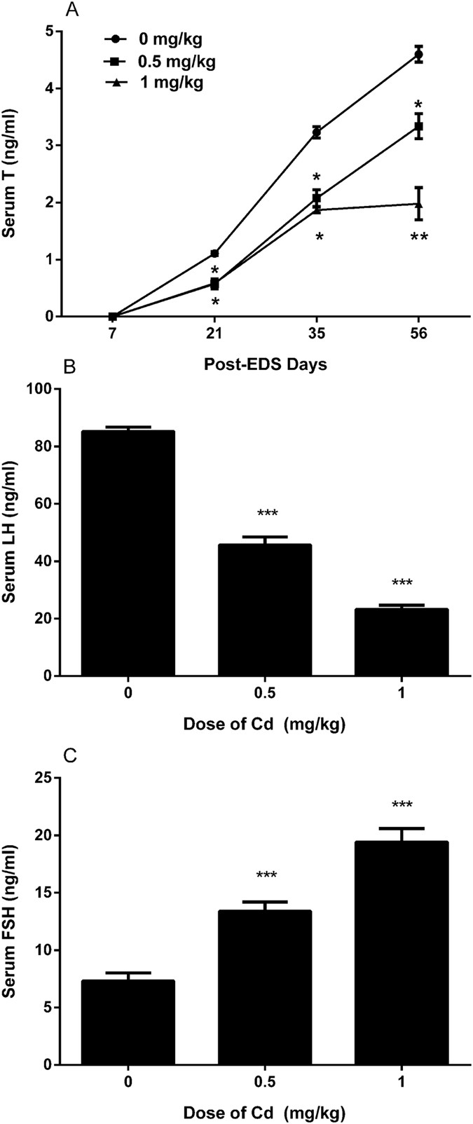
A brief exposure to cadmium impairs Leydig cell regeneration in the adult rat testis

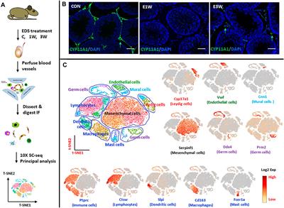
Cell Type-Specific Expression of Testis Elevated Genes Based on Transcriptomics and Antibody-Based Proteomics

Morphology of Leydig cells in the testes after in vivo MCP-1 treatment.

Antioxidants, Free Full-Text

Stem Leydig cells: Current research and future prospects of regenerative medicine of male reproductive health - ScienceDirect

The Sertoli cell: one hundred fifty years of beauty and plasticity - França - 2016 - Andrology - Wiley Online Library

Fluoride-Induced Autophagy via the Regulation of Phosphorylation of Mammalian Targets of Rapamycin in Mice Leydig Cells
Frontiers Identification of Rat Testicular Leydig Precursor Cells by Single-Cell-RNA-Sequence Analysis
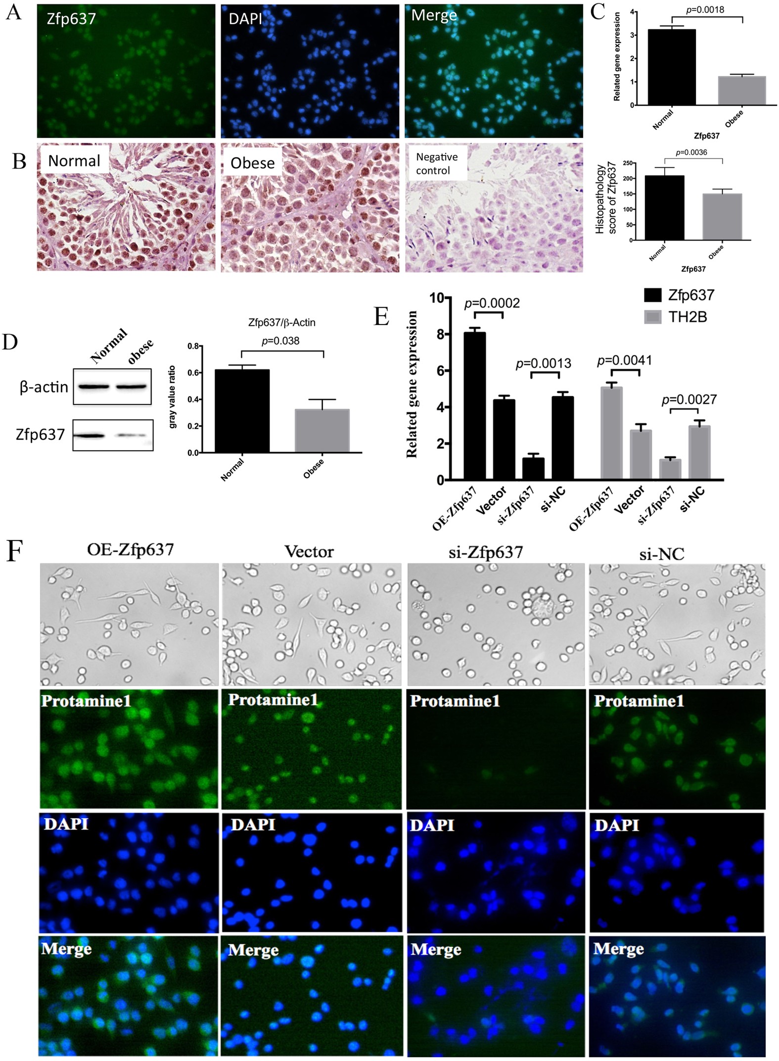
IL-6 mediates differentiation disorder during spermatogenesis in obesity-associated inflammation by affecting the expression of Zfp637 through the SOCS3/STAT3 pathway

Prenatal exposure to bisphenol AF induced male offspring reproductive dysfunction by triggering testicular innate and adaptive immune responses - ScienceDirect
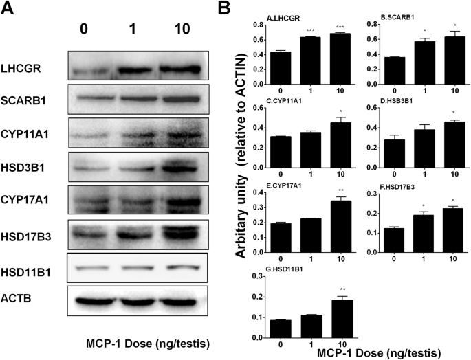
Monocyte Chemoattractant Protein-1 stimulates the differentiation of rat stem and progenitor Leydig cells during regeneration, BMC Developmental Biology

Testicular inflammation and infertility: Could chlamydial infections be contributing? - Bryan - 2020 - American Journal of Reproductive Immunology - Wiley Online Library
de
por adulto (o preço varia de acordo com o tamanho do grupo)