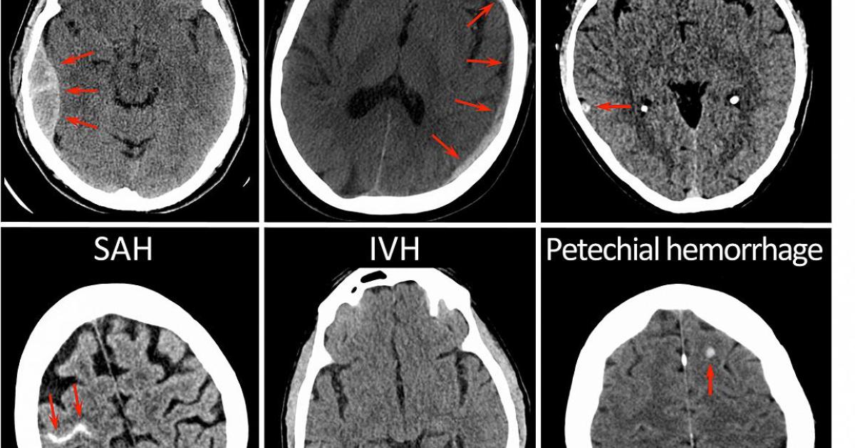
CT brain image gallery - SAH
Por um escritor misterioso
Descrição
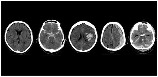
CT brain images - example of subarachnoid haemorrhage as seen on CT. Dense material in the basal cisterns, fissures and sulci represents acute bleeding into the subarachnoid space.

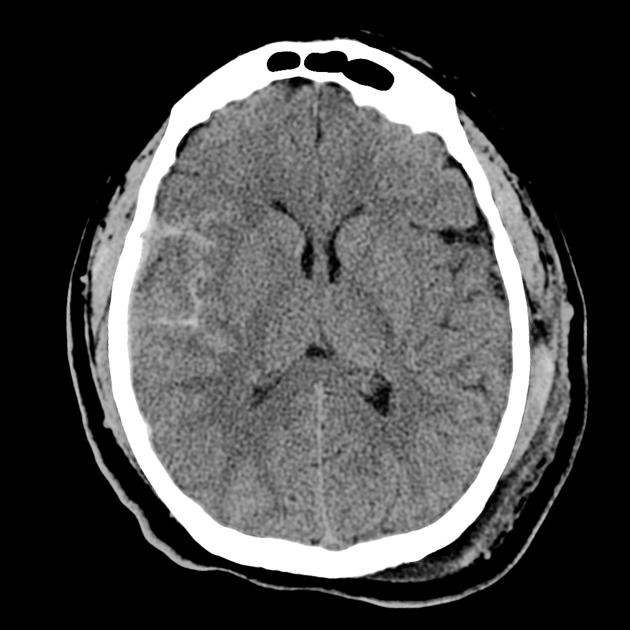
Typical CT scan showing subarachnoid haemorrhage. The image is

CT Angiography and Perfusion CT in Cerebral Vasospasm after
Sensors, Free Full-Text
Single Phase Dual-energy CT Angiography: One-stop-shop Tool for

Detection of hyperacute subarachnoid hemorrhage of the brain by
New CT patterns provide 'encouraging view' of traumatic brain

Research – Vascular Neurology Research Laboratory
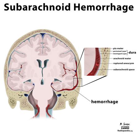
Subarachnoid hemorrhage Radiology Reference Article
Subarachnoid Haemorrhage (SAH), Overview

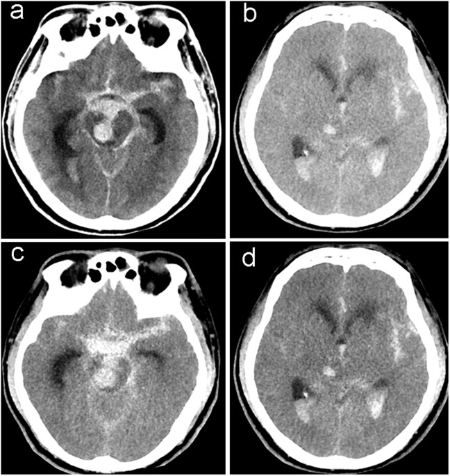
Axial brain CT scans obtained on admission, showing an SAH with
de
por adulto (o preço varia de acordo com o tamanho do grupo)